DYNAMICAL CELL SYSTEMS GROUP
Research

Dynamical Cell Systems Group

Research

Research

Research programmes

Our research integrates high‑content imaging, functional genetics and computational modelling to answer systems questions about shape, signalling and cell fate in cancer.
01

How do signalling networks encode and control cell shape?

Cell shape is both an output and an input to signalling networks. We investigate how biochemical pathways and mechanical cues are integrated to produce distinct morphological states and how those states influence downstream decisions in cancer cells.

Cell morphology reflects underlying signalling states: by quantifying shape we can infer pathway activity and regulatory interactions at scale. We use multiparametric single‑cell measurements from high‑content imaging combined with functional perturbations to reconstruct phenotype‑to‑network maps.

We study how changes in extracellular matrix, adhesion and cytoskeletal regulators reconfigure signalling modules (for example RAP1 and NF‑κB) and bias cells toward migratory, proliferative or drug‑tolerant states. Translating these mappings to human cancer datasets helps prioritise candidate regulators for functional validation.
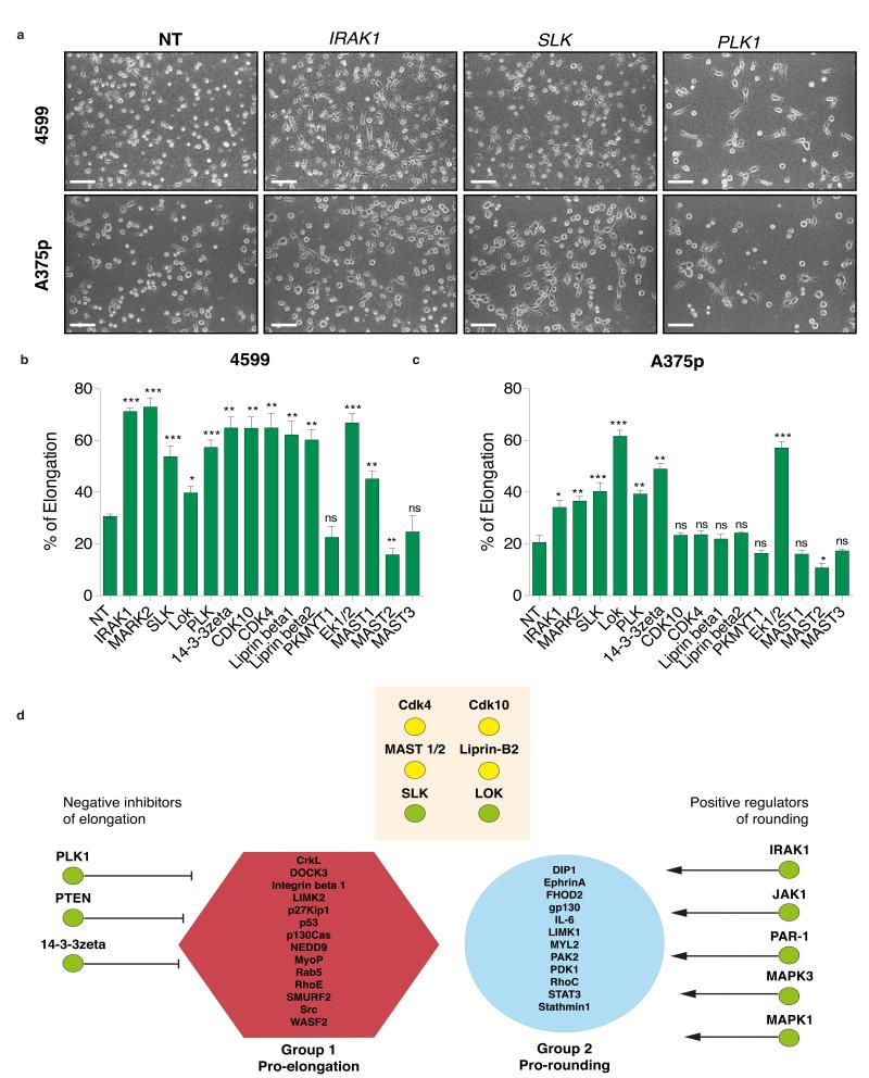

Representative morphological states from a high‑content screen that identified regulators of discrete cell shapes. (Adapted from Yin et al., Nat Cell Biol. 2013; Fig. 1.)
02

Discovery with high‑content, image‑based genetic screens

Automated microscopy and large‑scale perturbations allow systematic mapping of gene function in the context of morphology, cell cycle and signalling dynamics.

We design image‑based screens using Drosophila and human cell systems to detect phenotype changes across thousands of perturbations. By combining morphological readouts with network inference algorithms we reveal modules that coordinate cytokinesis, polarity and migration.

Our work emphasises scalable, reproducible pipelines for image acquisition, segmentation and feature extraction; these are coupled with statistical models that infer genetic relationships and signalling architecture from multiparametric phenotype space.

Figure 3 from 'A screen for morphological complexity identifies regulators of switch-like transitions between discrete cell shapes' (Nat Cell Biol, 2013) — morphology phenocluster analysis and representative cell-shape phenotypes used in high-content image-based genetic screens.
03

How cell‑cycle state gates signalling responses and therapy outcomes

Cell‑cycle position profoundly affects signalling dynamics and drug response. We interrogate how oscillatory programmes and state‑dependent signalling shape sensitivity to targeted therapies.

Using live‑cell imaging time series and latent‑state modelling we map how morphological dynamics and cell‑cycle progression interact with ERK/MAPK and other pathways. Temporal analysis reveals that signalling responses to BRAF/MEK inhibition are cell‑cycle dependent and can govern transient tolerance.

We develop hidden Markov models and related time‑series frameworks to describe transitions between morphological and cell‑cycle states, enabling prediction of when cells are most vulnerable to perturbation and how synchrony influences population responses.

Figure 1 (overview/representative images) from 'Receptor-Driven ERK Pulses Reconfigure MAPK Signaling and Enable Persistence of Drug-Adapted BRAF-Mutant Melanoma Cells' (Cell Systems, 2020) — illustrative ERK-KTR live-cell reporter images and schematics showing pulsatile ERK signalling relevant to cell-cycle–dependent drug responses.
04

Mechanisms that determine cancer cell fitness under stress

We study the determinants of cancer cell survival and fitness under genetic, metabolic and drug‑induced stress, focusing on links between morphology, signalling rewiring and adaptive responses.

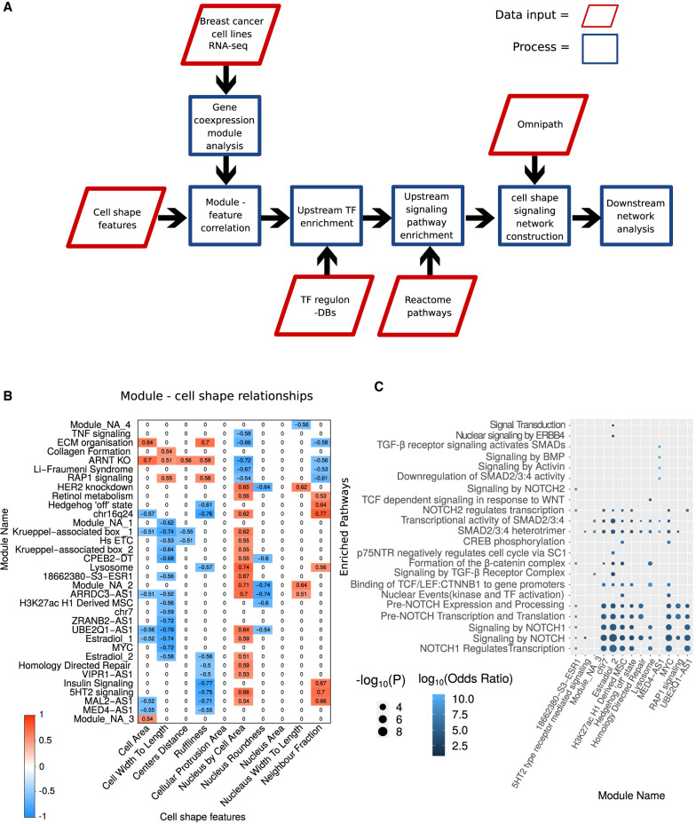
By integrating transcriptomic, proteomic and image‑derived features we identify phenotype‑specific modules—such as RAP1 signalling and NF‑κB activation—that mediate responses to mechanical and biochemical stress.

Our translational aim is to map conserved fitness programmes across tumour types and prioritise candidate regulators whose modulation sensitises cells to therapy or blocks metastatic competence.

Integration of gene expression modules with image‑derived morphological states highlights phenotype‑specific signalling networks linked to cancer cell fitness. (Adapted from Sero et al., Genome Biol. 2022.)